Diagnostics & Testing
Our team at Mount Sinai Medical Center is here to help you move towards accurate diagnosis of your cardiovascular condition – and to find the most effective treatment possible. Our leading-edge echocardiography laboratory at the Columbia University Division of Cardiology at Mount Sinai is accredited by the Intersocietal Commission for the Accreditation of Echocardiography Laboratories (ICAEL) and is recognized as one of the best in the region.
Our team of experts – all board-certified in cardiology and echocardiography – utilizes the latest technology, including 3D imaging, to make even the most difficult diagnoses via noninvasive techniques. We perform more than 6,000 transthoracic and 1,500 transesophageal studies annually, which places us among the most active heart institutes in the nation.
Angiography
Angiography (also known as an angiogram) is an X-ray of the blood vessels (arteries or veins) in the body. An angiogram uses a contrast medium, or dye, which shows up on the X-ray to help your Mount Sinai physician identify problems within your heart. In addition to the heart, this technique can be used to look at many areas of the body, including the brain, neck, heart, chest and lungs, limbs, and kidneys.
At the Mount Sinai Heart Institute, we perform angiography in our state-of-the-art catheterization lab, or cath lab for short. During an angiogram, a long, thin tube or catheter is inserted into a large artery. At Mount Sinai, we frequently use the radial artery in the wrist, but sometime use an artery in the groin area. Then, the catheter is threaded through the artery until its tip reaches the blood vessel your doctor needs to evaluate. A small amount of dye is then injected into the blood vessel through the catheter, and X-ray pictures are taken. Then a Mount Sinai specialist studies the X-rays to identify any potential issues.
Angiography can be used to diagnose a number of cardiovascular conditions, including aneurysms, arterial stenosis, malformations of the heart, and clots.
Non-Invasive Testing
Non-invasive cardiac testing does not require catheters to be inserted into the heart. Still, these tests can be used to detect serious heart conditions, including coronary heart disease, or plaque in the arteries, and coronary defects. These tests substantially improve our ability at Mount Sinai to accurately diagnose heart conditions without the need for invasive procedures.
Advanced Lipid Profile
Your Mount Sinai cardiologist may order a lipid profile to better determine your risk for coronary heart disease. An advanced lipid profile test measures your cholesterol levels – both good and bad cholesterol – in the blood and is a good indicator of whether someone may have a blockage of blood vessels or hardening of the arteries, which increase the overall risk of heart attack or stroke.
Cholesterol is a lipid (or fat) that is carried through the heart and body in the bloodstream and is either produced by the liver or consumed through food. Your cholesterol level is determined by a combination of genetics and diet, and having an unhealthy level can put you at risk for heart disease, stroke, and other diseases.
A standard cholesterol test measures your LDL level (or “bad” cholesterol), HDL level (or “good” cholesterol), triglycerides, and total cholesterol. An advanced lipid profile, however, analyzes the makeup of LDL and HDL cholesterol to measure the subtypes of each kind. While a standard cholesterol test can help gauge your risk of developing cardiovascular disease, an advanced lipid profile test can improve the accuracy of the test from around 40 percent to around 90 percent.
Ankle Brachial Index (ABI)
ABI compares blood pressure at the ankle to blood pressure in the arm, and at the Mount Sinai Heart Institute, we use the ABI to diagnose Peripheral Artery Disease (PAD). When someone’s blood pressure in the legs is less than 90% of the blood pressure measured in the arms, it can indicate the vessels in the legs are blocked, which means they may have PAD.
Cardiac Calcium Scoring
A cardiac calcium score test, also known as a heart scan, measures the amount of plaque buildup in the coronary arteries. Calcium buildup found in the arteries is a sign of coronary heart disease. At Mount Sinai, we perform a CT scan, which uses computers and a rotating machine to take X-ray images of the body from different angles to create a three-dimensional look at your heart and the arteries in the heart. If your Mount Sinai physician detects large amounts of plaque, further testing, including an angiography, may be necessary.
Echo Stress Test
The echo stress test is a supplement to the routine exercise cardiac stress test. During stress echocardiography, sound waves (ultrasound) are used to produce images of the heart at rest and at the peak of exercise.
EKG (Electrocardiography)
An EKG, which is shorthand for electrocardiography, is a commonly used heart test that primarily provides your Mount Sinai cardiovascular specialist with information about the heart’s rhythms. It is often used in combination with an echocardiogram. In an EKG, we place sticky pads on the chest, which leads to the EKG machine. These leads transmit information about how the heart is beating, based on its electrical activity. The lines created from one beat to the next create a pattern that helps your Mount Sinai doctor easily identify if something may be wrong with your heartbeat or detect signs of blockages or of a heart attack.
Exercise Stress Test
While it may sound stressful, a cardiac stress test is a non-invasive test to show your Mount Sinai cardiac care team how your heart is working during physical activity. This test, also called a treadmill test or exercise test, is performed while you’re walking on a treadmill or pedaling on an exercise bicycle. During the test, you’ll be connected to an electrocardiogram, or ECG, which monitors your heart rhythm. At Mount Sinai, we also measure your blood pressure during a stress test to get a better picture of the impact physical activity has on your heart and circulatory system.
Holter Monitor
The Holter monitor is a small, portable electrocardiogram (ECG) machine. The device is worn in a pouch around the neck or waist, and it keeps a record of the patient’s heart rhythm, typically over a 24-hour period. Your Mount Sinai cardiovascular expert will usually ask you to keep a diary that records your activity during the 24-hour period, along with any symptoms you feel, such as chest pain, fatigue, dizziness, or palpitations. Then, using the results from the monitor, along with your diary, we’re able to better understand the impact your activity has on your heart.
Nuclear Stress Test
At Mount Sinai, we use nuclear stress tests to get a more thorough picture of your heart and vessels than can be seen during a regular exercise stress test. During a nuclear stress test, we measure blood flow to the heart muscle at rest and during stress by having you perform activity, like walking on a treadmill. Prior to the test, we’ll inject a radioactive tracer that shows up on certain types of advanced imaging devices that we use at Mount Sinai, including position emission tomography (PET) and single photo emission computed tomography (SPECT). These devices take pictures of your heart that give us a detailed view of how your heart looks during the stress of physical exertion.
Imaging
The Mount Sinai Heart Institute is home to advanced imaging technology that allows your cardiovascular care team to view both your heart’s function and its anatomy (or structure). Our imaging capabilities not only provide valuable diagnostic information to better evaluate your heart health and plan your treatment, but in many cases, our imaging studies are completely non-invasive.
Cardiac MRI (CMR)
Cardiovascular magnetic resonance imaging (CMR), also known as a cardiac MRI, is a medical imaging technology for the non-invasive assessment of the function and structure of the cardiovascular system. It uses the same basic principles as magnetic resonance imaging (MRI), but we optimize the imaging to better evaluate the cardiovascular system. This provides in-depth information that can diagnose and evaluate a multitude of conditions, evaluating the function of the heart, its valves, and heart chamber size.
Carotid Ultrasound
A carotid ultrasound is a non-invasive ultrasound method used to examine blood circulation. Our team takes an ultrasound of the body’s two carotid arteries, which are located on each side of the neck and carry blood from the heart to the brain. The carotid ultrasound provides detailed pictures of these blood vessels and information about the blood flowing through them. A carotid ultrasound can include two different types of ultrasounds:
- Doppler Carotid Ultrasound – This shows your Mount Sinai cardiovascular care team how blood flows through the carotid arteries.
- Standard Carotid Ultrasound – This shows us images of the structure of the carotid arteries.
Chest X-Ray
A chest X-ray is a radiology test that produces images of the heart, blood vessels, lungs, and airways. It also produces images of the bones of the chest and spine. For this test, the chest is exposed briefly to radiation, which produces an image of the chest, as well as the internal organs in the chest. A chest X-ray can show the following inside the body:
- Calcium deposits
- Fractures
- Heart-related lung concerns
- Lung conditions
- Medical device placement
- Shape and size of the heart
Computerized Tomography (CT) Coronary Angiogram
Unlike a conventional angiogram that requires a catheter to be inserted in the heart, a computerized tomography (CT) coronary angiogram is a non-invasive test. At Mount Sinai, we use CT coronary angiogram to view the arteries that supply blood to the heart. The CT coronary angiogram uses a sophisticated type of X-ray that produce images of the heart and blood vessels in three dimensions. This is a valuable tool in the diagnosis of coronary artery disease, and because it is non-invasive, it does not require recovery time and has almost zero risk.
Echocardiogram
An echocardiogram is a test that uses sound waves, or ultrasound, to produce images of your heart. The echocardiogram shows your Mount Sinai cardiovascular care team how the heart muscle and valves are working. If their view of your heart is blocked by your lungs or ribs, we may decide to in inject an enhancing agent to make the pictures of the heart’s structures appear more clearly on the monitor.
Our Physicians
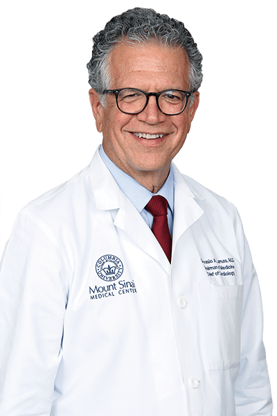
Gervasio A Lamas, MD
Chairman of Medicine
Eugene J. Sayfie Chair of Cardiology
Chief, Columbia University Division of Cardiology at Mount Sinai Medical Center
Co-Director, Mount Sinai Heart Institute
Professor at the Columbia University Division of Cardiology at Mount Sinai Medical Center
- Cardiology
- Concierge Medicine
- Mount Sinai Medical Center (Main Campus)
- 305.674.2998
Alfonso O Tolentino, MD
Vice Chairman of Medicine
Director of Electrocardiography Laboratory at the Columbia Division of Cardiology at Mount Sinai Medical Center
Assistant Professor of Medicine at the Columbia University Division of Cardiology at Mount Sinai Medical Center
- Cardiology
- Mount Sinai Medical Center (Main Campus)
- 305.674.2690
Roy F Williams, MD
Chief, Divison of Thoracic Surgery
- Cardiology
- Robotic Surgery
- Thoracic & Cardiovascular Surgery
- Lung Cancer
- Mount Sinai Medical Center (Main Campus)
- 305.674.2121
Nirat Beohar, MD
Director, Cardiac Catheterization Laboratory
Director, Interventional Cardiology Fellowship Program
Medical Director, Structural Heart Disease Program
Professor at the Columbia University Division of Cardiology at Mount Sinai Medical Center
- Interventional Cardiology
- Cardiology
- Mount Sinai Medical Center (Main Campus)
- 305.674.2690
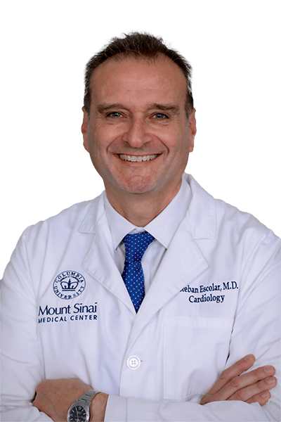
Esteban Escolar, MD
Director, Coronary Care Unit
Director, Cardiology Clinical Services
Program Director, Brodie Family Cardiovascular Disease Fellowship
Associate Professor at the Columbia University Division of Cardiology at Mount Sinai Medical Center
- Cardiology
- Mount Sinai Medical Center (Main Campus)
- 305.674.2121
- Mount Sinai Primary & Specialty Care Skylake
- 305.919.1970
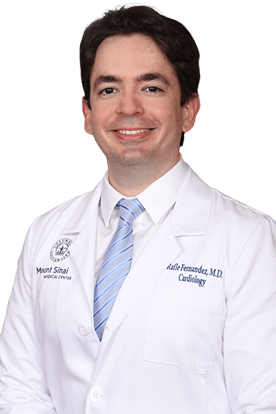
Rafle Fernandez, MD, MBA, FACC
Associate Director, Clinical Echocardiography
Assistant Professor of Medicine at the Columbia University Division of Cardiology at Mount Sinai Medical Center
- Cardiology
- Mount Sinai Primary & Specialty Care Miami Shores
- 305.891.3710


