The diagnosis and treatment of heart disease has undergone significant changes over the past fifty years. Landmark clinical trials performed over this period of time, coupled with a dramatic increase in technological advances have allowed us to better understand the number one killer in the USA.
Here’s what you need to know about the three most common, non-invasive imaging tools used to help with the diagnosis of heart disease:
Echocardiography
- Most common imaging tool used in cardiology
- Has been around since the 1960s
- Noninvasive – Uses sound waves to create a moving picture of the heart and blood flow within it
- A small probe is placed on the chest of the patient by a technologist
- Images are interpreted by a highly-trained cardiologist (at Mount Sinai, cardiologists are Echo Board Certified)
- It usually takes about 30 minutes
- It’s painless and has no side effects

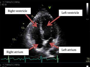
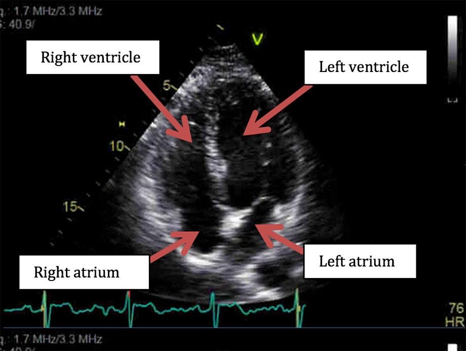
(A still frame of a patient’s heart obtained with echocardiography.)
Computed Tomography (CT)
- Latest available technology that can take images of the beating heart, and show calcium deposits and blockages in your heart arteries
- Noninvasive, uses X-rays
- Two types of CT: one type requires an IV to administer iodine contrast dye. The other does not require contrast dye. Both options give patients and physicians different types of information.
- Scan takes between 5-10 minutes
- Coronary Computed Tomography Angiography (CCTA) allows for the creation of three-dimensional images of the coronary arteries. Helps determine if plaque buildup has narrowed the blood vessels that supply the heart.

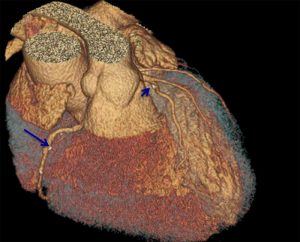
(Example of what a CCTA looks like in a patient with a small amount of plaque buildup in the right coronary artery (arrow) and a left anterior descending artery)
Magnetic Resonance Imaging (MRI)
- More sophisticated imaging exam yields images based on the effect of a powerful magnet
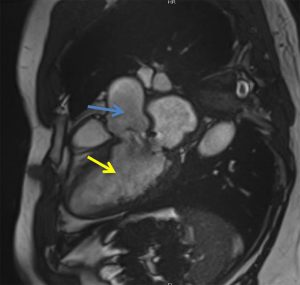
- Used to detect or monitor cardiac disease and evaluate the heart’s anatomy
- Does not use X-rays radiation
- Test may take up to 60- 90 minutes
Now you have a basic understanding of the differences between these three non-invasive imaging tools, which help us diagnose heart disease. However, it is important for you to realize that clinical history and physical examination remain the most critical tools to diagnose heart disease. Finally, the interpretation of images requires expertise available at Mount Sinai.


