- Color Doppler & Duplex Studies
- Echocardiography
- Transcranial Doppler
Radiology (Imaging) Services Overview
Specialty Team of Radiologists
Mount Sinai offers the only on-site board certified radiology subspecialist team on Miami Beach. This high degree of training ensures that each patient will get the most accurate diagnosis. Professional interpretation of your study is vital to not only accurately diagnose your condition, but also to help plan any needed treatment. In addition, as a teaching hospital our faculty radiologists keep current on the latest imaging techniques which also helps to best determine your diagnosis.
Radiation Safety Provider
Mount Sinai outpatient imaging is a Radiation Safety Provider which means we have instituted safeguards to ensure the smallest amount of radiation dose exposure to patients undergoing testing. These safeguards include:
- ensuring you receive the correct diagnostic study required for the suspect diagnosis,
- using proper techniques and protocols to obtain the information needed with the least amount of exposure,
- eliminating duplicative studies, and
- maintaining previous imaging studies for reference.
Secure Electronic Systems
The testing results are available electronically to your physician immediately upon dictation. Our radiologists also personally call your physician with any critical findings. Your imaging history is maintained in Mount Sinai’s secure electronic patient network that your physician can see from his/her computer. Electronic record storage also allows our radiologists to access your history with each new exam to detect changes in your condition. This can helps avoid unnecessary radiation exposure by eliminating duplicative studies that do not contribute to your diagnosis.
Hospital Quality Care and Services
Using hospital-based outpatient imaging services provides many benefits, including:
- Quality results interpreted by board certified specialty trained radiologists
- High quality state-of-the-art equipment
- Advanced imaging, data storage, and communications technology
- Comprehensive imaging modalities in one location
- Secure patient history for reliable personalized results
- Diagnostic and treatment coordination through one quality provider
- Stable, reliable, accredited services
- Flexible locations and flexible scheduling
- Oversight of radiation safety practices
Imaging Services
Mount Sinai Medical Center’s Lila and Harold Menowitz Comprehensive Breast Center was the first breast center in Florida to be accredited by the National Accreditation Program for Breast Centers (NAPBC). This accreditation is given only to those centers that have passed a rigorous evaluation, are committed to provide the highest quality care in breast disease diagnosis, treatment, support and follow-up care and have demonstrated they can comply with the accreditation program’s high standards.
DEFINITION: Computerized tomography (CT) scanning combines special X-ray equipment with sophisticated computers to produce multiple images of the inside of the body. A series of X-rays are taken from many different angles that produce cross-sectional images of your bones and soft tissues. CT scan images provide much more information than plain X-rays and are an efficient, painless exam.
WHAT YOU CAN EXPECT: Before your CT, you may be asked to remove your clothing and change into a hospital gown. You will need to remove any metal objects, such as jewelry, that might interfere with imaging. CT scanners are shaped like a large doughnut standing on its side. You will be asked to lie on a narrow table that slides into the “doughnut hole” or gantry. As the X-ray tube rotates around your body, the table slowly moves through the gantry. While the table is moving, you may need to hold your breath to avoid blurring the images.
A technologist will be watching you from the next room and you will be able to communicate with him or her via intercom, should you need any assistance during the scan. CT scans only take a few minutes.
HOW TO PREPARE: To properly visualize some areas, you may need to fast for a period of time beforehand or receive contrast orally or intravenously. Movement blurs the images and may lead to inaccurate results. Ask your doctor how best to prepare if you think you may have issues remaining still during the exam.
- IF YOUR SCAN REQUIRES CONTRAST: All CT patients scheduled for abdomen, pelvis, adrenals, liver, retro, pancreas or kidney scans must drink oral contrast as follows: by 8 p.m. the night before the appointment (if the appointment is scheduled before noon the following day) or by 7 a.m. the same day (if the appointment is scheduled after noon). Also, the patient must not eat or drink for four hours prior to the procedure.
- Do not drink oral contrast for CT kidney scans if you have a diagnosis of kidney stones.
- The oral contrast prep can be picked up Monday – Friday 8 a.m. to 6 p.m. at both of our diagnostic centers or at your physician’s office during business hours.
- Patients scheduled for CT enterography (images of the small bowel) must not eat or drink anything, including medications, for four hours prior to the procedure. Patients must report to the diagnostic center at least one hour prior to the appointment time. At this time, they will be given 16 ounces of water and a contrast agent to drink over the course of the hour before the test. Patients may empty their bladder prior to the scan.
- Patients scheduled for CT program (images of the urinary tract) must not eat or drink anything after midnight the night before the scan.
RESULTS: One of our specially trained radiologists will review the images from your scan and generate a report of any findings. Either the doctor who performed your test or the doctor who asked you to have a CT angiogram will discuss the results of the test with you.
DEFINITION: A CT simulation for radiation therapy follows your initial consultation in our Radiation Therapy Department. CT simulation includes a CT scan of the area of your body to be treated with radiation. The CT images acquired during your scan will be reconstructed and used to design the best and most precise treatment plan for you. The simulation portion of your radiation therapy regiment ensures that your treatments will target the area of concern, while missing surrounding critical structures.
WHAT TO EXPECT: You will begin the simulation by having a CT scan of the area of your body to be treated with radiation therapy. We will ensure that you are as comfortable as possible for your scan, as the goal is to position you the same way every day during your treatment regimen. The very center of the treatment area will be defined and marked as a reference point to be used during your treatment. We may need to use permanent ink to create a very small tattoo in this area. This will be discussed with you before simulation and only takes a couple of seconds.
The second half of simulation involves the creation of an immobilization device that will be utilized over the duration of your treatment to keep you in the same position each and every time you are treated. A mask may be used for treatments near the head, and a “cradle” or “mold” may be cast around the area of interest if it is located on the torso or body . Marks will be made on the mask or mold to serve as reference points that help our radiation therapists place you in the correct and exact position every day, ensuring your treatments are as accurate and precise as possible.
HOWTO PREPARE: Your radiation oncologist will give you specific instructions on how to prepare for your CT simulation during your initial consult. Simulation can sometimes be scheduled the next day, depending on availability.
WHAT HAPPENS AFTER SIMULATION: The images obtained during your CT simulation will be used to plan your treatments. Depending on the type of treatment you will receive and the various tasks associated with it, you will begin radiation therapy 1-10 days after initial simulation.
A Doppler ultrasound test uses reflected sound waves to see how blood flows through a blood vessel. It will assist your doctor in evaluating blood flow through major arteries and veins, such as those of the arms, legs and neck. It can reveal blocked or reduced blood flow that may cause a stroke. It also can reveal blood clots in leg veins that may break loose and block blood flow to the lungs. (During pregnancy, Doppler ultrasound may be used to look at blood flow in an unborn baby to check the health of the fetus. Duplex Doppler ultrasound uses standard ultrasound methods to produce a picture of a blood vessel and the surrounding organs. The computer then converts the Doppler sounds into a graph that provides additional information about the speed and direction of blood flow through the blood vessel being evaluated.
WHAT TO EXPECT: For ultrasound testing, gel is applied to the skin to help transmit the sound waves during the exam. A small, handheld instrument called a transducer is passed back and forth over the area of the body that requires viewing. The transducer sends out high-pitched sound waves < above the range of human hearing ) that are reflected back to the transducer. A computer analyzes the reflected sound waves and converts them into an image that is displayed to the technologist.
HOW TO PREPARE: You may need to stop using products that contain nicotine ( cigarettes or chewing tobacco ) between 30 minutes and two hours before the test. Nicotine causes blood vessels to constrict and may give false results.
Patients scheduled for gallbladder, liver, pancreas, abdominal aorta, renal arteries and SMA/celiac ultrasounds should not have anything to eat or drink from midnight the night before the appointment.
RESULTS: One of our specially trained radiologists will analyze the images from your scan and report the findings to your referring physician or primary care doctor. Your doctor will then discuss any important findings and next steps with you.
DEFINITION: An echocardiogram uses sound waves to produce images of your heart. This test is commonly used to see how your heart is beating and pumping blood to identify various abnormalities in the heart muscle and valves.
WHAT TO EXPECT: You will be asked to undress from the waist up and change into a hospital gown. Before you start the test, stickers/patches (electrodes) will be placed on your chest. You will then be asked to lie on an examining table or bed for the exam.
If you will have a transesophageal echocardiogram, your throat will be numbed with a numbing spray or gel, and you will likely be given a sedative to help you relax.
During the echocardiogram, the technician will dim the lights to better view the images on the monitor. You may hear a pulsing sound while the machine records the blood flow through your heart. Sometimes the transducer must be held very firmly against your chest to help the technician produce the best images of your heart. Our technologist will take special care to make sure you are as comfortable as possible during the procedure. You may be asked to breathe in a certain way or to roll onto your left side in order to obtain necessary images.
Most echocardiograms take less than an hour, but the timing may vary depending on how many images need to be taken.
HOW TO PREPARE: No special preparations are necessary for a standard transthoracic echocardiogram. Your doctor will ask you not to eat for a few hours beforehand if you’re having a transesophageal or stress echocardiogram. If you’re having a transesophageal echocardiogram, be sure to make arrangements for someone to drive you home because of the sedating medication you will receive.
RESULTS: One of our specially trained radiologists will interpret your results. Depending on your results, you may be referred to a heart specialist (cardiologist) for more tests. Treatment depends on what’s found during the exam and your specific signs and symptoms. You may need a repeat echocardiogram in several months or other diagnostic tests, such as a cardiac computerized tomography (CT) scan or CT angiogram.
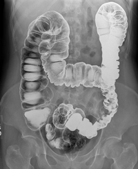 DEFINITION: Fluoroscopy is a non-invasive exam that is utilized to visualize your gastrointestinal tract in motion. Fluoroscopy uses a continuous X-ray beam to create a sequence of live images that are projected onto a television-like monitor and interpreted by one of our specially trained radiologists (link to “meet our Radiologists”). A contrast material, which appears bright white on the image, clearly defines the area being examined and makes it possible for a physician to view internal organs in motion. Still images are also captured and stored digitally on a computer.
WHAT TO EXPECT: The technologist will explain your procedure and answer any questions that you may have prior to your exam. You will then be asked to remove all clothing and change into a gown. Several exams can be conducted under fluoroscopy. The following are the most common and what you can expect during each exam.
UPPER GI SERIES
For the first part of the exam, the table is in the upright position. You will be standing on a platform attached to the table. The radiologist will instruct you to take a sip of barium (contrast) from the cup the technologist has provided. Next, you will be given a cup containing “crystals” that the technologist will add water to and ask you to drink it as quickly as possible. These crystals create gas in the stomach, which aids in the imaging of the esophagus and stomach. It is important that you try not to belch. The radiologist will watch under X-ray as you swallow the barium and take several small images of various portions of the esophagus and stomach as you drink.
You will then be asked to lie on the table for the second part of the exam. The technologist will then ask you to take several sips of barium through a straw. The radiologist will record images as the barium makes its way to the stomach. When the radiologist is finished viewing this process, the technologist will take images of the stomach in various positions. TOTAL TIME REQUIRED: Approximately 45 minutes to an hour.
HOW TO PREPARE: Patient must not eat or drink after midnight the night before the test.
ESOPHAGRAM
For the first part of the esophagram, the table is in the upright position. You will be standing on a platform attached to the table. The radiologist will instruct you to take a sip of barium from the cup the technologist has provided. Next, you will be given a cup containing “crystals” that the technologist will add water to and ask you to drink it as quickly as possible. These crystals create gas in the stomach, which aids in the imaging of the esophagus and stomach. It is important that you try not to belch. The radiologist will then watch as you swallow the barium and take several small images of various portions of the esophagus and stomach as you drink.
For the second part of the esophagram, you will lie flat on the table. You will then be asked to take several sips of barium through a straw. The radiologist will record images as the barium makes its way to the stomach. When the radiologist is finished viewing this process, the test is complete.
TOTAL TIME REQUIRED: Approximately 30-45 minutes.
HOW TO PREPARE: No specific preparation is required.
SMALL BOWEL SERIES
You will be asked to lie on the table on your back to begin the exam. An image will be taken to make sure that your intestines are clean. The purpose of the test is to fill the small intestine with barium (contrast). This means that you may need to consume several cups of barium. We need you to drink as much as possible, but will only ask you to drink as much as you can tolerate. After you finish drinking the liquid, you will be asked to lie on your right side to help move the liquid from your stomach into your small intestine.
The normal “transit time” through the small bowel varies widely, and may be between 30 minutes and one hour. Several X-rays will be taken in 15 minute intervals until the barium reaches its destination. The images are then reviewed by the radiologist, who may need to take additional images. Once the barium has passed through the small intestine and the images are satisfactory to the radiologist, the test is complete.
TOTAL TIME REQUIRED: The test can takes between 30 minutes and several hours, depending on how fast the liquid moves through your intestines.
HOW TO PREPARE: Patient must not eat or drink after midnight the night before the test.
BARIUM ENEMA
This exam is used to visualize your lower intestine and requires contrast administered through your rectum. First, you will be asked to lie on the x-ray table on your side. The technologist will then gently insert a small lubricated enema tip into your rectum by which the barium will be administered. As the barium enters the colon, you may feel some pressure along with some cramping. The barium will flow outlining your colon as the radiologist will ask you to hold your breath while he or she records images. Slight pressure may be applied to your abdomen to help the barium outline the colon. After this portion of the exam is completed, the technologist will take additional images. Once the films are complete you will be able to go to the bathroom and empty your colon. One additional image will be taken afterwards.
TOTAL TIME REQUIRED: Approximately 45 minutes.
HOW TO PREPARE: Please pick up prep from your physician or as instructed. Patient must ONLY drink clear liquids the day before the test, and must not have anything to eat or drink after midnight the night before the test. RESULTS: Our specially trained radiologists will interpret the images taken during your scan and report the results to your referring physician.
We look forward to seeing you and will be more than happy to answer any other questions you may before, during and after your procedure.
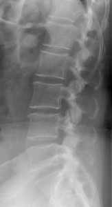 DEFINITION: An X-ray is a quick, painless test that produces images of the structures inside your body — particularly your bones. X-ray beams can pass through your body, but they are absorbed in different amounts depending on the density of the material they pass through. Dense materials, such as bone and metal, show up as white on X-rays. The air in your lungs shows up as black. Fat and muscle appear as varying shades of gray.
For some types of X-ray tests, a contrast medium — such as iodine or barium — is introduced into your body to provide greater detail on the X-ray images.
WHAT TO EXPECT: You may be asked to change into a gown depending on the area of your body being X-rayed. A technologist will position your body to obtain the necessary views. He or she may use pillows or sandbags to help you into the proper position. During X-ray exposure, you will be asked to remain still and hold your breath. Any movement can blur the image.
An X-ray procedure may take only a few minutes for a bone X-ray, or more than an hour for moreinvolved procedures, such as those done under fluoroscopy.
Your child’s X-ray
If a young child is having an X-ray, immobilization techniques may be used to help keep him or her still. These will not harm your child and will prevent the need for a repeat procedure. You may be allowed to remain with your child during the test, but will be asked to wear a lead apron to shield you from unnecessary exposure.
After the X-ray
After an X-ray, you generally can resume normal activities. Routine X-rays usually have no side effects.
HOW TO PREPARE: Different types of X-rays require different preparations. Ask your doctor or nurse to provide you with specific instructions.
Contrast material
Before some types of X-rays, you will be given a liquid called contrast medium. Contrast mediums, such as barium and iodine, help outline a specific area of your body on the X-ray image. You may swallow the contrast medium, or receive it as an injection or an enema.
Barium enema – Please pick up prep from your physician or as instructed. Patient must ONLY drink clear liquids the day before the test, and must not have anything to eat or drink after midnight the night before the test.
- IVP – Please pick up prep from your physicianor as instructed. Patient must not have anything to eat or drink after midnight the night before the test.
- Upper GI Series – Patient must not have anything to eat or drink after midnight the night before the test.
- Small Bowel Series – Patient must not have anything to eat or drink after midnight the night before the test.
- Gastrogaffin GI – Patient must not have anything to eat or drink after midnight the night before the test.
- Barium Swallow-Esophagram – No prep required.
- CT/IVP Combo – Please pick up prep from your physician or as instructed. Patient must not have anything to eat or drink after midnight the night before the test.
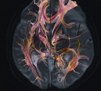 High field Magnetic Resonance Imaging (MRI) is one form of imaging used by physicians to obtain clinically useful diagnostic information. Incorporating advanced technology, it produces images of anatomy without the use of radiation required with other imaging modalities, such as X-ray or computed tomography (CT). MRI combines the physical properties of strong magnetic fields with radio waves to produce computer-generated soft tissue images within any plane of the body. This popular imaging technique can be used as a primary diagnostic tool to provide a quick and accurate diagnosis for your physician. In some situations, this procedure can reduce the need for further diagnostic procedures or invasive procedures, such as exploratory surgery, that may have associated complications. At Mount Sinai, we utilize the latest technology in MRI with high field equipment to ensure the clearest image possible for your exam.
Specially trained interventional radiologists use imaging guidance such as ultrasound, x-ray, computed tomography (CT) or magnetic resonance imaging (MRI) to perform minimally invasive procedures…
DEFINITION: A magnetic resonance angiogram (MRA) is an imaging test used to look at the arteries that supply blood to your heart muscle. MRAs don’t require the recovery time needed with traditional angiograms. Unlike a traditional coronary angiogram, an MRA relies on a powerful MRI machine to produce images of your heart and heart vessels. In many cases, MRA can provide information that can’t be obtained from an X-ray, ultrasound, or computed tomography (CT) scan.
An MRA is commonly performed to look for:
- A bulge (aneurysm), clot or the buildup of fat and calcium deposits (stenosis caused by plaque) in the blood vessels leading to the brain
- An aneurysm or tear (dissection) in the aorta, which carries blood from the heart to the rest of the body
- Narrowing (stenosis) of the blood vessels leading to the lungs, kidneys or legs
WHAT TO EXPECT: You will be asked to change into a gown and to remove jewelry, hairpins eyeglasses, watches, wigs, dentures, hearing aids and underwire bras, as these items are a safety concern while around magnetic equipment.
For your scan, you will be asked to lie down on a movable table that slides into the opening of the tunnel. During the scan, the internal part of the magnet produces repetitive tapping and other noises. Earplugsor music may be provided to help block the noise. You will be instructed to breathe normally but to lie as still as possible, as movement can blur the resulting images. A technologist will monitor you closely from another room and you will be able to communicate with him or her, should you require immediate assistance. An MRA typically lasts less than an hour.
The technician might ask you to hold your breath for 10-15 seconds at a time while taking pictures of your heart. A contrast agent, such as gadolinium, might be used to highlight your blood vessels or heart in the pictures.
IF YOUR EXAM REQUIRES CONTRAST: Contrast will be administered to you during your scan. You may feel a cool sensation during the injection and discomfort when the needle is inserted. Gadolinium, the contrast most often used, does not contain iodine.
HOW TO PREPARE: Before an MRA scan, eat normally and continue to take your usual medications, unless otherwise instructed. If you are worried about feeling claustrophobic during the scan, talk to your doctor beforehand, as he or she may be able to prescribe medication to ease your anxiety.
RESULTS: One of our specially trained radiologists will analyze the images from your scan and report the findings to your referring physician or primary care doctor. Your doctor will then discuss any important findings and next steps with you.
DEFINITION: Magnetic resonance imaging (MRI) is used to create multi-dimensional pictures of your brain in order to look for differences in structure. MRI spectroscopy is used to specifically look at the chemical make-up of different brain regions.
WHAT TO EXPECT: You will be asked to change into a gown and to remove all jewelry, hairpins eyeglasses, watches, wigs, dentures, hearing aids and underwire bras, as these items are a safety concern while around magnetic equipment.
For your scan, you lie down on a movable table that slides into the opening of the tunnel. A technologist monitors you closely from another room and you will be able to communicate with them via intercom. During the MRI scan, the internal part of the magnet produces repetitive tapping and other noises. Earplugs or music may be provided to help block the noise. You must hold very still because movement can blur the resulting images. If you are worried about feeling claustrophobic inside the MRI machine, talk to your doctor beforehand. An MRI scan typically lasts less than an hour. Gathering information for the spectroscopy portion of the exam may require an additional 10 to 15 minutes.
HOW TO PREPARE: Before an MRI scan, eat normally and continue to take your usual medications, unless otherwise instructed.
RESULTS: One of our specially trained radiologists will analyze the images from your scan and report the findings to your referring physician or primary care doctor. Your doctor will then discuss any important findings and next steps with you.
DEFINITION: Magnetic resonance imaging (MRI) is a test that uses a magnetic field and pulses of radio wave energy to make pictures of structures inside the body. The MRI machine looks like a tunnel with two open ends. In some cases, contrast material may be used during the MRI scan to show certain structures more clearly.
WHAT TO EXPECT: You will be asked to change into a gown and to remove all jewelry, hairpins eyeglasses, watches, wigs, dentures, hearing aids and underwire bras, as these items are a safety concern while around magnetic equipment.
For your scan, you lie down on a movable table that slides into the opening of the scanner. A contrast agent (dye) may be injected through an intravenous (IV) line in your arm to enhance the appearance of tissues or blood vessels on the MRI pictures. During the scan, the internal part of the magnet produces repetitive tapping and other noises. Earplugs or music may be provided to help block the noise. You will be instructed to breathe normally but to lie as still as possible, as movement can blur the resulting images. A technologist monitors you closely from another room and you will be able to communicate with them via intercom, should you need assistance during the scan. An MRI typically lasts less than an hour.
HOW TO PREPARE: Before an MRI scan, eat normally and continue to take your usual medications, unless otherwise instructed. If you are worried about feeling claustrophobic during the scan, talk to your doctor beforehand, as he or she may be able to prescribe medication to help ease your anxiety.
RESULTS: One of our specially trained radiologists will analyze the images from your scan and report the findings to your referring physician or primary care doctor. Your doctor will then discuss any important findings and next steps with you.
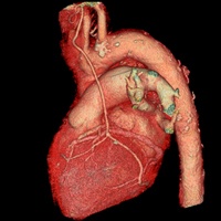 Multi-slice CT (Computerized Tomography) imaging is a quick and painless scan that allows your doctor to see greater detail than plain X-rays. It uses X-ray images from many different angles to create cross-sectional images of bones and soft tissue for diagnostic and treatment purposes. The biggest advantage of using multi-slice CT is that it can image various different parts of the body fairly quickly and can be used various capacities, from trauma to treatment planning for radiation therapy. It can also serve as an alternative to invasive surgical procedures.
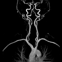 Mount Sinai’s non-invasive vascular lab uses Doppler ultrasound techniques to examine patients without the risks and discomforts associated with injections and other invasive procedures. Almost all known or suspected vascular disorders can be tested and diagnosed by this ultrasound technique. Testing can also often determine the severity of a patient’s vascular problems and theneed for treatment.
Non-invasive Vascular Lab Services include:
DEFINITION: Open magnetic resonance imaging (MRI) uses MRI technology but offers many benefits for claustrophobic, bariatric, geriatric, orthopaedic and pediatric patients. Mount Sinai offers the largest MRI machine opening on the market for your comfort. MRI is a test that uses a magnetic field and pulses of radio wave energy to make pictures of structures inside the body. The open MRI machine looks different than a traditional MRI machine because it offers a large opening with nonobstructed side views for your ease and comfort. As with traditional MRI scans, contrast material (dye) may be used during open MRI scans to show certain structures more clearly.
There are many benefits of using our open MRI unit:
- Patients will have a clear 270° view for virtually every exam without compromising the quality or efficiency of your study.
- Open views reduce anxiety in claustrophobic patients and decrease their need for sedation medication during the test.
- The table can accommodate up to 660 lbs and lower to wheelchair height for easy access.
- The large opening accommodates patients who have broader shoulders and do not fit into a traditional MRI unit. It allows allows for easier positioning of limbs during all exams.
- The large opening enables parents of children who are being imaged to have constant contact with their child during the test.
Please schedule your visit at our Aventura location to receive the highest quality open MRI services available.
WHAT TO EXPECT: You will be asked to change into a gown and to remove all jewelry, hairpins eyeglasses, watches, wigs, dentures, hearing aids and underwire bras, as these items are a safety concern while around magnetic equipment. For your scan, you lie down on a movable table that slides into the open scanner. A contrast agent (dye) may be injected through an intravenous (IV) line in your arm to enhance the appearance of tissues or blood vessels on the MRI pictures. During the scan, the internal part of the magnet produces repetitive tapping and other noises. Earplugs or music may be provided to help block the noise. You will be instructed to breathe normally but to lie as still as possible, as movement can blur the resulting images. A technologist monitors you closely from another room and you will be able to communicate with them via intercom, should you need assistance during the scan. An MRI typically lasts less than an hour.
HOW TO PREPARE: Before an MRI scan, eat normally and continue to take your usual medications, unless otherwise instructed. If you are worried about feeling claustrophobic during the scan, talk to your doctor beforehand, as he or she may be able to prescribe medication to ease your anxiety.
RESULTS: One of our specially trained radiologists will analyze the images from your scan and report the findings to your referring physician or primary care doctor. Your doctor will then discuss any important findings and next steps with you.
DEFINITION: Diagnostic ultrasound, also called sonography or diagnostic medical sonography, is an imaging method that uses high-frequency sound waves to produce relatively precise images of structures within your body. It does not use X-rays or any other type of possibly harmful radiation and can be used for diagnosing and treating a variety of diseases and conditions.
A Doppler ultrasound test uses reflected sound waves to see how blood flows through a blood vessel. It will assist your doctor in evaluating blood flow through major arteries and veins, such as those of the arms, legs and neck. It can reveal blocked or reduced blood flow through narrowing in major arteries of the neck that may cause a stroke. It can also be used to evaluate blood flow after a stroke or other condition that might be caused by a problem with blood flow. Evaluation of a stroke can be done through a technique called transcranial Doppler (TCD) ultrasound, which evaluates the blood flow of the brain.
WHAT TO EXPECT: For ultrasound testing, gel is applied to the skin to help transmit the sound waves during the exam. A small, handheld instrument called a transducer is passed back and forth over the area of the body that is of interest. The transducer sends out high-pitched sound waves < above the range of human hearing > that are reflected back to the transducer. A computer analyzes the reflected sound waves and converts them into an image that is displayed to the technologist.
HOW TO PREPARE: You may need to stop using products that contain nicotine < cigarettes, chewing tobacco > for 30 minutes to 2 hours before the test. Nicotine causes blood vessels to constrict and may give false results.
RESULTS: One of our specially trained radiologists will analyze the images from your scan and report the findings to your referring physician or primary care doctor. Your doctor will then discuss any important findings and next steps with you.
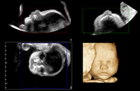 DEFINITION: Ultrasound, also called sonography or diagnostic medical sonography, is an imaging method that uses high-frequency sound waves to produce relatively precise images of structures within your body. It does not use X-rays or any other type of possibly harmful radiation and can be used for diagnosing and treating a variety of diseases and conditions.
Most ultrasound examinations are done using a sonar device outside of your body, though some ultrasound examinations involve placing a device inside your body.
WHAT TO EXPECT: For ultrasound testing, a gel is applied to the skin to help transmit the sound waves during the exam. A small, handheld instrument called a transducer is passed back and forth over the area of the body that is of interest. The transducer sends out high-pitched sound waves (above the range of human hearing) that are reflected back to the transducer. A computer analyzes the reflected sound waves and converts them into a picture that is displayed to the technologist. The picture produced by ultrasound is called a sonogram or ultrasound scan.
Though the majority of ultrasound exams are performed with a transducer on your skin, some ultrasounds are done inside your body (invasive ultrasounds). For these exams, a specialized transducer is attached to a probe that’s inserted into a natural opening in your body. Most commonly, this is exam is a transvaginal ultrasound, which is inserted into a woman’s vagina.
Ultrasound is usually a painless procedure. However, you may experience some mild discomfort as the sonographer guides the transducer over your body, especially if you’re required to have a full bladder. Our technologist will take special care to make sure you are as comfortable as possible during the scan. A typical ultrasound exam takes from 30 minutes to an hour.
HOW TO PREPARE: How you prepare for an ultrasound depends on the area of interest you are having imaged:
- Patients scheduled for gallbladder, liver, pancreas, abdominal aorta, renal arteries and SMA/celia ultrasounds should not have anything to eat or drink after midnight the night before the appointment.
- Patients scheduled for pelvis/bladder ultrasound must drink 32 ounces of water or juice 30 -45 minutes prior to the scheduled appointment time.
- Patients scheduled for obstetrics ultrasounds do not need to drink water prior to procedure.
RESULTS: One of our specially trained radiologists will analyze the images from your scan and report the findings to your referring physician or primary care doctor. Your doctor will then discuss any important findings and next steps with you.
DEFINITION: Virtual colonography is an exam used to detect changes or abnormalities in the large intestine (colon) and rectum. Unlike traditional colonoscopy, virtual colonography doesn’t require sedation or the insertion of a scope into the colon, which allows the patient to recover and resume normal activities more quickly. Virtual colonography uses an imaging technique known as computerized tomography (CT) to produce hundreds of cross-sectional images of the abdominal organs. The images are combined and digitally manipulated to provide a detailed view of the inside of the colon and rectum.
WHAT YOU CAN EXPECT: Before your virtual colonography, you will be asked to change into a gown. You’ll begin the exam lying on your side on the exam table, usually with your knees drawn toward your chest. The doctor will place a small tube (catheter) inside your rectum to fill your colon with air or carbon dioxide. The air or gas may cause a feeling of pressure in your abdomen. During virtual colonography, you may be given an injection of medication to reduce the likelihood of stomach cramps during the exam.
For the next part of the exam, you will be asked to lie on your back. The exam table will be moved into the CT machine, and your body will be scanned. You will be asked to lie on your abdomen or your side, and your body will be scanned again. You may be asked to turn and hold various other positions, as well as hold your breath, so that proper images can be acquired.
A virtual colonography typically takes about 10 minutes. After the exam, most of the air or gas will be removed from your colon through the catheter in your rectum. You may feel bloated or pass gas for a few hours after the exam as you clear the remaining air or gas from your colon. Walking may help relieve any discomfort, and you can return to your usual diet and daily activities immediately.
HOW TO PREPARE: Preparation for this exam is key to helping our doctors identify any suspicious areas in your colon or rectum. Before a virtual colonography, you’ll need to clean out (empty) your colon by following a strict diet.
- Twenty four hours before your procedure, begin a liquid diet (i.e. clear soup, water, gelatin, apple juice or any clear juice but NO MILK OR MILK PRODUCTS). You may drink plenty of clear liquids throughout the day.
- Prior to your procedure, pick up a prep kit from your physician or as instructed.
- At 10 a.m. the day before your procedure, begin using the prep kit as instructed.
- On the day of the procedure, have absolutely nothing to eat or drink, including medications. Remind your doctor of your medications at least a week before the exam. You may need to temporarily stop taking certain medications days or hours before the exam.
RESULTS: One of our specially trained radiologists will review the images from your scan and generate a report of any findings. Your doctor will then review the results of the colonography with you. A negative result means the doctor did not find any abnormalities in the colon. A positive result means images revealed polyps or other abnormal tissue in the colon.
Our Physicians
Chetan Rajadhyaksha, MD
Chairman, Department of Radiology
Chief, Section of Nuclear Imaging
- Radiology
- Nuclear Imaging
- Mount Sinai Medical Center (Main Campus)
- 305.674.2121
William F Burke III, MD
Chief, Section of Thoracic Imaging
Vice Chairman, Department of Radiology
Program Director, Radiology Residency Program
- Radiology
- Mount Sinai Medical Center (Main Campus)
- 305.674.2121
Vinay Bhatia, MD
Section Chief Neuroradiology
Director of MRI Services
- Neuroscience
- Neuroradiology
- Radiology
- Mount Sinai Medical Center (Main Campus)
- 305.674.2121
Stuart S. Kaplan, MD
Chief, Section of Breast Imaging, Breast Ultrasound and MRI, and Breast Interventional Procedures
- Cancer
- Oncology
- Radiology
- Breast Imaging
- Mount Sinai Medical Center (Main Campus)
- 305.674.2121
- Mount Sinai Medical Center (Main Campus)
- 305.932.4766
- Mount Sinai Emergency Center, Physician Offices, Cancer Center and Diagnostic Center Aventura
- 305.692.1010
Marcia C. Javitt, MD, FACR, FSAR, FSRU
Director of Radiology Research and Scholarly Services
- Radiology
- Mount Sinai Medical Center (Main Campus)
- 305.674.2121


